Plenary speakers deliver 45 minute lectures during the mornings of the scientific SSNR program. The lectures will span broad surveys of recent major developments in the neurorehabilitation field.
More content coming soon, please subscribe to our SSNR mailing list to be alerted on updates.

Northwestern University
Abstract: As a rehabilitation scientist, we share the dream of unraveling recovery mechanisms, developing new clinical or experimental tools, and ultimately helping more people. After years in this field, I find myself continuously cycling through literature review, hypothesis formulation, experimentation, data analysis, and clinical practice. Fortunately, over the past 20 years, I have made a spiral ascent in this cycle, culminating in leading a team that affirms my status as an independent researcher. Here, ‘independent’ means that my team and I can manage every aspect of this process. In this chain, PhD students make significant contributions. In return, PhD training provides the most structured way to equip a young person with practice-based learning opportunities to experience most, if not all, of these roles. In this talk, I aim to share my research experiences and insights on how to initiate and sustain this cycle effectively, and I extend my best wishes for you to achieve independence in your research career.
Dr. Jun Yao, with expertise in biomedical signal processing, neuroscience, and rehabilitation engineering, currently holds the position of Director at the Neuro-Image and Interfaces Laboratory. She is a full professor in both the Department of Physical Therapy and Human Movement Sciences at the Feinberg School of Medicine and the Department of Biomedical Engineering at the McCormick School of Engineering, Northwestern University. Dr. Yao’s team is dedicated to developing new methods that deepen our understanding of the neuro-mechanisms controlling movement in healthy individuals and the disorders that arise following a stroke. With these enhanced scientific insights, the team advances biomedical engineering techniques, such as neural machine interfaces, to restore functional control of the impaired hand in individuals with moderate to severe stroke impairment.
National Center for Adaptive Neurotechnologies
Albany Stratton VA Medical Center and State University of New York
Albany, New York, USA
Abstract: Once considered a backwater, neurorehabilitation is now among the most vibrant areas of preclinical and clinical biomedical research. Until recently, its main strategy has been skill-specific practice, which often fails to produce adequate recovery. Now, new recognition that the CNS remains plastic through life, new understanding of the CNS substrates of skilled behaviors, and newly developed technologies combine to redefine the therapeutic goal and provide new strategies that promise to improve the efficacy of skill-specific practice.
The substrate of a skill is a network of neurons and synapses that may extend from cortex to spinal cord. This network has been given the name heksor, based on the ancient Greek word hexis. Each heksor changes through life; it modifies itself to maintain the key features of its skill, the attributes that make the skill satisfactory. Muscle activity and kinematics may change; key features are maintained. Heksors overlap; they share CNS neurons and synapses. Through their concurrent changes, they keep CNS neuronal and synaptic properties in a negotiated equilibrium that enables each heksor to achieve the key features of its skill.
When CNS damage disrupts important skills, the primary therapeutic goal is to enable damaged heksors to repair themselves and reestablish a negotiated equilibrium in which each can once again produce its skill satisfactorily. Two new strategies can increase the efficacy of skill specific practice. One new strategy increases the capacity for plasticity. This gives damaged heksors more options for self-repair: they shape the additional plasticity through skill-specific practice. The other new strategy targets beneficial plasticity to a critical site in a damaged heksor. This improves skill-specific practice, which enables the heksor to achieve much wider beneficial plasticity. In animals and humans, combining skill-specific practice with these strategies produces large clinically significant improvements in recovery. These improvements persist.
For basic and clinical researchers, the challenge is to develop effective multitherapy protocols – protocols that combine these new strategies with skill-specific practice. Computational modeling can help identify and parameterize promising protocols. Controlled clinical trials that evaluate a new protocol mechanistically and compare it to current state-of-the-art treatment are essential. Assessments should evaluate relevant skills, overall function, and quality of life, and should extend at least six months after therapy ends. Study of pre-morbid factors as well as reflexes, evoked potentials, muscle activity, and kinematics during treatment can guide patient specific protocol modifications. Many multitherapy protocols will be noninvasive and suitable for home use with remote oversight. Reference: Wolpaw JR, Kamesar A. Heksor: the central nervous system substrate of an adaptive behaviour. Journal of Physiology 600.15:3423-3452, 2022. DOI: 10.1113/JP283291
Dr. Wolpaw is a neurologist who has devoted nearly 50 years to basic and clinical research. His group developed operant conditioning of spinal reflexes as a model for defining the plasticity underlying learning, and went on to show that this conditioning can improve walking in animals and people with spinal cord injuries. This work introduced the new therapeutic method of targeted neuroplasticity. His group also guided development of brain-computer interface (BCI) principles and methods and is exploring BCI use to improve neurorehabilitation. Most recently, in response to the growing appreciation of the lifelong plasticity of the CNS, he led formulation of a new paradigm for understanding how useful behaviors are acquired and maintained through life, a paradigm based on the new concepts of heksors and the negotiated equilibrium of CNS properties that heksors create. His group has been supported throughout by NIH, the VA, and private foundations; their work has been recognized by many national and international awards.
The Hong Kong University of Science and Technology, Hong Kong
Abstract: The ability to learn and adapt to the environment is a fundamental aspect of biological behavior, which has been achieved through remarkable mechanisms developed by the brain. To enable the brain to control neuroprosthetic devices via brain-machine interfaces (BMI), it is critical to model and recreate such adaptive mechanisms. Coadaptive BMIs offer a promising solution for paralyzed individuals by allowing subjects and external devices to adapt to each other during closed-loop control. However, current systems still face challenges such as extensive training, frequent recalibration, and lack of generalization over new tasks. An ideal BMI should assist subject learning with proper feedback, achieve high decoding accuracy and stability, and improve task learning efficiency over time.
To address these limitations in BMI systems, we designed a framework that facilitates subject learning on new tasks with dynamic sensory feedback and improves decoders’ learning ability through kernel reinforcement learning (KRL) methods. Our coadaptive framework involves an additional closed-loop that incorporates medial prefrontal cortex (mPFC) activity. The neural dynamics of audio-induced mPFC activity are translated into reward expectation information to guide the learning of decoders on a new task.
Our results demonstrate that the coadaptive framework improves the behavioral learning of subjects and the decoder’s learning efficiency on new tasks. The framework advances BMIs into a more adaptive and efficient system with learning ability on new tasks. Our study has the potential to empower clinical BMI devices with learning, where the subject learns how to execute complex movements through daily interactions.
Yiwen Wang received her B.S. and M.S. degrees from University of Science and Technology of China (USTC), Hefei, Anhui, China respectively. She received a Ph.D. degree from University of Florida, Gainesville, FL, USA. She joined as an associate professor at Zhejiang University, Hangzhou, China. She is now an associate professor with substantiation at the Department of Electronic and Computer Engineering, Department of Chemical and Biological Engineering, the Hong Kong University of Science and Technology.
Her research interests are in neural decoding of brain-machine interfaces, adaptive signal processing, computational neuroscience, and neuromorphic engineering. She serves as the Chair of the IEEE EMBS Neural Engineering Tech Committee, the chair of the IEEE BRAIN publication subcommittee, and the board member of Brain Computer Interfaces Society. She is the Editor-in-Chief of the IEEE Brain Newsletter. She also serves on the editorial board of the Journal of Neural Engineering, and is the associate editor of Frontiers in Human Neuroscience (Brain-Computer Interfaces), an associate editor of the IEEE Transactions on Neural Systems and Rehabilitation Engineering, and an associate editor of the IEEE Transactions on Cognitive and Developmental Engineering. She was recognized as IEEE EMBS distinguished lecturer in 2022, and received IEEE EMBS distinguished service award in 2023. She holds two US patents and has authored more than 100 peer-reviewed publications.
Instituto de Biomecánica de Valencia, Spain
In collaboration with Juan-Manuel Belda-Lois
Abstract: We’ll present the results of a series of experiments conducted to explore new avenues to the analysis of human movements through the scanning of shapes in motion, also known as 4D scanning. 4D scans can provide trajectories of points on the surface of human bodies, with unique identifiers that can be associated with anatomical landmarks without having to instrument the participants. It has been proved that those data can be used to analyze the kinematics of movement, with differences from marker-based systems comparable to inter-examiner errors. In meshes with nearly 50,000 vertices, it is possible to represent anatomical landmarks using dense distributions of points over the target body regions and perform an analysis of gait kinematics with Bayesian statistics. Other ways of measuring the orientation of body segments are possible without having to identify specific landmarks that may be difficult to locate, such as tracking the rotation of contours of body sections with characteristic shapes.
4D scans can be also the source of metrics based on the shape and topology of body segments. Body segment inertial parameters, measured directly from the geometry of the segmented mesh at each instant, can be used to improve joint moment estimation through inverse dynamic analysis in situations where the inertial effect of body masses is significant, in neuromusculoskeletal models this implies reduction of the residuals. Kinematic alterations and asymmetric patterns in soft tissue deformation between the right and left legs have also been found at different instants of the gait cycle, and related to differences in muscle activation, consistent with clinical assessments of people with neurological disorders.
Dr. Rosa Porcar-Seder holds a Ph.D. in Industrial Engineering from the Universitat Politècnica de València. She has been working at the Instituto de Biomecánica de Valencia since 1995. First as a researcher, then as a business intelligence manager. Her main research topics were occupational biomechanics, human factors applied to the automotive industry, and head-eye mobility patterns related to progressive lens design strategies. Now, she is focused on technology transfer, promoting the research and development of use cases of biomechanical technology to solve new problems in industry and health.
ETH Zurich, Switzerland
Abstract: Rehabilitation robots can be very useful to restore movement abilities in two ways. First, they can promote neurorehabilitation as training devices after neurological injuries such as spinal cord injury (SCI), traumatic brain injury and stroke. Second, they can be used as assistive devices to support patients or elderly people with motor impairments in daily life situations. However, current solutions are still too bulky, too heavy and with too little battery power. Furthermore, joint misalignments often lead to mechanical stress in the attachment points, and patient engagement and motivation is limited. These disadvantages result in discomfort and unsatisfactory performance. This talk will present new solutions and current trends of stationary movement training robots as well as wearable exoskeleton devices that are more convenient than previous devices and provide patient-cooperative control strategies in order to better engage the patient during the movement. A new soft, lightweight, and under-actuated tendon-driven exo-muscular system makes the technology wearable so that it provides more mobility during therapeutic training in hospitals and higher comfort and performance for daily use in home and work environments. Different systems have been developed for the support of the lower extremities (“MyoSuit”) and the upper extremities (“MyoShirt”). They are currently tested in many patients with different kinds of muscle weaknesses, e.g. paraparesis after SCI, hemiparesis after stroke, Multiple Sclerosis, different kinds of muscle dystrophies, and patients with heart failure.
Robert Riener studied Mechanical Engineering at TU München, Germany, and University of Maryland, USA. He received a Dr.-Ing. degree in Engineering from the TU München in 1997. After postdoctoral work at Politecnico di Milano and TU München he became assistant professor at ETH Zurich and Spinal Cord Injury Center of the University Hospital Balgrist (“double-professorship”) in 2003; since 2010 he has been full professor for Sensory-Motor Systems at ETH Zurich, and since 2016 also full professor at the medical faculty of the University of Zurich. He was guest professor at USC Los Angeles, SJTU Shanghai, and SSSA Pisa. Riener has published more than 500 peer-reviewed journal and conference articles, 36 books and book chapters and filed 28 patents. Riener is the initiator and organizer of the CYBATHLON, which was honored with the European Excellence Award, the Yahoo Sports Technology Award and two REIMAGINE Education Awards. In 2018 Riener obtained the honorary doctoral degree from the University of Basel. Since 2019, Riener is AAAS Leshner Leadership Fellow and since 2022 he is the president of the ICORR (International Consortium of Rehabilitation Robotics).
ETH Zurich, Switzerland
Abstract: Advances in peripheral nervous system (PNS) interfacing present a promising venue for rehabilitation of individuals with different neurological disabilities. Subjects with diabetes or lower-limb amputation frequently do not engage fully in everyday activities because they are afraid of falls. They also tend to have reduced mobility, which can induce a sedentary lifestyle that promotes disease development and hinders reinsertion into society, while the neuropathic pain is also common and poorly managed with current medications. Despite a wide range of possibilities for human-machine interfacing, the nature of the optimal human-machine interaction remains poorly understood. Knowledge gained from in-silico modelling of targeted neural structures can inform an optimized design of such interfacing, therefore we develop the exact models of different nerves, enabling for AI-based personalized treatments. We have pioneered a human-machine systems that translates prosthetic sensors’ read-outs into “language” understandable by the nervous system. A “sensing leg,” for lower- limb amputees, by connecting sensors from the prosthetic knee and under the foot to the residual PNS, transduces the readout of the sensors into stimulation parameters. The “smart orthosis” for diabetics “speaks” to their residual healthy nerves with a similar philosophy. Their effects at the brain level were evaluated, observing important benefits. These studies not only provided clear evidence of the benefit of neuromodulation for neurologically disabled subjects but also provided insights into fundamental mechanisms of supraspinal integration of the restored sensory modalities.
Stanisa Raspopovic has been an Assistant Professor of Neuroengineering at the Swiss Federal Institute of Technology, Zurich, Switzerland and is an incoming Full Professor of Biomedical Engineering. His research interest is focused on the development of innovative medical devices for treatment of neurologically disabled persons. In particular, he develops mechatronic systems directly interfacing the environment with the residual nervous system. Stanisa achieved the groundbreaking translational research results in the field of sensory restoration in amputee patients. He has substantial international experience in research in neural engineering culminating with the award of an ERC Starting grant in 2018, ERC Consolidator Grant in 2023, Science & PINS Prize in Neuromodulation 2021 and ETH Latsis Prize 2021.
Northwestern University, USA
Abstract: A recent survey of persons with high level spinal cord injury revealed that nearly 80% would undergo brain surgery to restore some use of their hands. To this end, the potential of existing intracortical Brain Computer Interfaces (iBCIs) lends real hope, yet even the most advanced iBCIs require the user to be wired to stationary equipment and allow only limited, intermittent control of a robotic hand. A number of years ago we developed a novel iBCI that used muscle activity (EMG) predictions to control the intensity of electrical stimulation of the temporarily paralyzed muscles of a monkey’s hand, so called, “Functional Electrical Stimulation” (FES). This FES iBCI allowed the monkeys to control not only the motion of their wrist and fingers, but also to exert force in a manner much more like the natural control of hand movement than with iBCIs that control only the kinematics of movement. Beyond the ability to restore voluntary limb movement, there is evidence that the tight synchrony between a user’s attempted movement and the peripheral stimulation that causes that movement, may invoke mechanisms of neural plasticity that could accelerate recovery from an actual spinal cord injury. In this talk I will describe the basic work that led to our proof-of-concept demonstration in monkeys, as well as our recent work to develop wireless versions of the FES iBCI that could allow restoration of a broader range of the activities of daily living. Finally, I will also describe our most recent efforts to develop related methods for humans with spinal cord injury.
For most of my academic career, I have done work that addresses basic properties of the neural control of reaching and grasping. My early training in Physics and Engineering has very much influenced my approach to these problems, which tends strongly to the quantitative. The recent growth of neural engineering has provided a productive avenue for the transformation of my basic research into a more applied, translational line of work. We have developed a very successful efferent interface that restores hand use to temporarily paralyzed monkeys using Functional Electrical Stimulation (FES) of the paralyzed muscles that is controlled in real time by motor cortical (M1) recordings. That project now uses wireless recording of both neural and EMG signals while the monkey is in its home cage, which affords us the unique opportunity to collect virtually unlimited datasets for decoder training during unconstrained, natural behavior. These datasets have allowed us to study the representation of hand use by M1 neurons across a wide range of natural behaviors, and to compare these representations to those of more typical constrained lab tasks. However, this particular model of SCI lacks critical elements, including muscle atrophy, denervation, and the opportunity for recovery of function through neuroplastic changes in spinal circuitry and in spared descending projections. For this reason, we have also developed a rodent model of cortically-controlled FES that we are using to restore locomotion in a rat with an actual lumbar hemisection. The project combines neuromuscular and spinal stimulation delivered wirelessly through an implanted device of our design.
Delft University of Technology, the Netherlands
University of Bern, Switzerland
Abstract: The recovery of sensorimotor functions after an acquired brain injury requires a long, highly intense, and repetitive training program. Several new technologies have been developed to support this highly demanding training, among the most prominent being robotic devices and virtual reality. Recently, there has been an increased interest in minimally supervised and unsupervised rehabilitation to increase therapy dosage in group therapy and complement conventional therapy at patients’ homes. These new inventions should be co-created between different stakeholders if we aim to facilitate their usability and acceptance by the final user. In this talk, I will present how we follow a human-centered design approach to design highly intuitive and usable haptic robotic devices for upper limb rehabilitation and immersive virtual reality environments using off-the-shelf Head Mounted Displays (HMD) to provide a rich high fidelity multisensory rehabilitation experience in minimally supervised environments.
Laura Marchal-Crespo is an Associate Professor at the Department of Cognitive Robotics, Faculty 3mE. She is also associated with Erasmus MC (Rotterdam) and the Faculty of Medicine at the University of Bern (Switzerland). Her research focuses on the general areas of human-machine interaction and biological learning and, in particular, the use of robotic devices and immersive virtual reality for the assessment and rehabilitation of patients with acquired brain injuries such as stroke. Webpage: www.mlnlab.nl
Kessler Foundation, USA
Abstract: To be announced
The Polytechnic University of Milan, Italy
Abstract: Nowadays, we are surrounded by digital tools and novel technologies. Are these technologies able to change the actual model of care providing valid information at the point of need?
In this lecture the methodology to co-design, develop and validate novel digital tools will be presented in general and in the specific use case of a smart ink pen to monitor handwriting. Handwriting represents a harmony of cognitive, sensory and fine motor control abilities. Thus, alterations in handwriting are indicative signs of an umbrella of neurological pathologies that concern both the motor and cognitive spheres. In this context a digital tool able to measure reliable and valid indicators of performance, coupled with an explainable machine learning algorithms can produce meaningful alerts at the point of need.
Simona Ferrante is an Associate Professor at the Electronics Information and Bioengineering Department, Politecnico di Milano. Her research activities are carried out at the Neuroengineering and Medical Robotics Laboratory (NearLab) in the field of neurorehabilitation, functional electrical stimulation and e-health. She is mainly focused on the development and validation of technologies for the rehabilitation, the transparent monitoring and the assessment of neurological patients and fragile people.
Imperial College London, United Kingdom
In collaboration with Oskar Aszmann and Antonio Bicchi
Professor Farina has been Full Professor at Aalborg University, Aalborg, Denmark, (until 2010) and at the University Medical Center Göttingen, Georg-August University, Germany, where he founded and directed the Institute of Neurorehabilitation Systems (2010-2016) until he moved to Imperial College London as Chair in Neurorehabilitation Engineering. His research focuses on biomedical signal processing, neurorehabilitation technology, and neural control of movement. Within these areas, he has (co)-authored approximately 400 papers in peer-reviewed Journals and >500 conference abstract and papers. He has been the President of the International Society of Electrophysiology and Kinesiology (ISEK) (2012-2014) and is currently the Editor-in-Chief of the official Journal of this Society, the Journal of Electromyography and Kinesiology. He is also currently an Editor for IEEE Transactions on Biomedical Engineering and the Journal of Physiology, and previously covered editorial roles in several other Journals.

University of Stanford, USA
Abstract: A publication is the principal output and measure of impact of work performed in universities. Scholarly publications frequently describe a discovery, invention, or novel method. In some instances, the positive impact of scholarly work on society can be greatly increased by translating the discovery, invention, or method into a product that is deployed on a scale that is much larger than is typical for a scholarly publication. This is especially important in biomedical research and engineering because of the potential for research to improve human health. In this lecture, I will describe three approaches that are effective for translating research into products that improve human health: a needs-driven approach, a technology-driven approach, and an open-source approach. I will use examples from my work in neuromodulation, neuromuscular physiology, and biomechanics to illustrate these approaches and make observations that apply more generally. My goal is to share insights that will help those who are interested in technology translation be successful in their endeavors.
Scott Delp, Ph.D., is the James H. Clark Professor of Bioengineering, Mechanical Engineering, and Orthopaedic Surgery at Stanford University. He is the Founding Chairman of the Department of Bioengineering at Stanford and Director of the Wu Tsai Human Performance Alliance, which aims to transform human health through the science of peak performance. Scott is also the Director of the RESTORE Center, a NIH national center focused on measuring real world rehabilitation outcomes and Director of the Mobilize Center, a NIH National Center of Excellence focused on Big Data and Digital Health. Scott’s laboratory develops technologies to advance movement science and human health. Software tools created in his lab, including OpenSim and Simtk.org, have become the basis of an international collaboration involving thousands of scientists who exchange simulations of human movement. He has published over 300 research articles and has recently released a book from MIT Press entitled Biomechanics of Movement: The Science of Sports, Robotics, and Rehabilitation. Professor Delp has co-founded six health technology companies and is a member of the U.S. National Academy of Engineering.
Volker Dietz
Balgrist University Hospital, Switzerland
Abstract: Rehabilitation should be designed on the basis of neurophysiological insights underlying normal and impaired sensorimotor functions, which requires interdisciplinary collaboration and background knowledge. Recovery of sensorimotor function after CNS damage is based on the exploitation of neuroplasticity, with a focus on the rehabilitation of movements needed for self-independence. This requires a physiological limb muscle activation that can be achieved through functional arm/hand and leg movement exercises and the activation of appropriate peripheral receptors. Such considerations have already led to the development of innovative rehabilitation robots with advanced interaction control schemes and the use of integrated sensors to continuously monitor and adapt the support to the actual state of patients. For a positive impact on outcome of function, rehabilitation approaches should be based on neurophysiological and clinical insights, keeping in mind that recovery of function is limited. When appropriately applied, robot-assisted therapy can provide a number of advantages over conventional approaches, including a standardized training environment, adaptable support and the ability to increase therapy intensity and dose, while reducing the physical burden on therapists. Rehabilitation robots are thus an ideal means to complement conventional therapy in the clinic, and bear great potential for continued therapy and assistance at home using simpler devices. In my talk I will highlight fundamental neurophysiological factors influencing the recovery of sensorimotor function after a stroke or spinal cord injury. I will thus provides insights on essential neurophysiological mechanisms to be considered for a successful rehabilitation.
Volker Dietz is Senior Researcher and emeritus Professor of the University of Zürich. His research is focused on human motor control and movement disorders using electrophysiological techniques. Major achievements were made by the introduction of technology in neurorehabilitation, such as the ‘Lokomat’ for the training of stepping movements and by establishing the neurophysiological basis of neurorehabilitation after stroke and spinal cord injury (SCI). Further major achievements were made in the field of spinal cord regeneration (completion of phase I trial using Nogo-A-antibodies; together with Novartis company and Martin Schwab) and in the field of exploitation of neuroplasticity by the activation of relevant receptors during training after CNS damage.
The Sant’Anna School of Advanced Studies, Italy
Abstract: Upper limb amputation deprives individuals of their innate ability to manipulate objects. Such disability can be restored with a robotic prosthesis linked to the brain by a human-machine interface (HMI) capable of decoding voluntary intentions, and sending motor commands to the prosthesis. Clinical or research HMIs rely on the interpretation of electrophysiological signals recorded from the muscles. However, the quest for an HMI that allows for arbitrary and physiologically appropriate control of dexterous prostheses, is far from being completed. Here we propose a new HMI that aims to track the muscles contractions with implanted permanent magnets, by means of magnetic field sensors. We called this a myokinetic control interface. In this talk I will present the concept, the features and the work done in the past years to prove its feasibility, limits and potentials in implementing direct and simultaneous control over multiple digits of an artificial hand.
Prof. Christian Cipriani is the Head of the Artificial Hands Area at the BioRobotics Institute of the Scuola Superiore Sant’Anna (SSSA) in Pisa, Italy. His field of research is (bio)mechatronics applied to the area of upper limb prosthetics. From 2017 to 2023 he served the BioRobotics Institute of SSSA as the Director. In 2012 he has been a Visiting Scientist with the Biomechatronics Development Lab at University of Colorado Denver | Anschutz Medical Campus, with a Fulbright Scholarship. He received the Ph.D. in Biorobotics Science and Engineering from a joint program between IMT Institute for Advanced Studies, Lucca and Scuola Superiore Sant’Anna in 2008, and the Laurea degree in Electronic Engineering (MSc) with full marks from the University of Pisa, Italy in 2004. He has coordinated several national and international research projects and authored 150+ peer reviewed scientific papers, 100+ of which on international journals. His current research is sponsored by the Italian Workers’ Compensation Authority (INAIL), the national Ministries of University and Research, Defence, Health, and the European Research Council (ERC). He is the founder of Prensilia S.r.l., a spin-off company of SSSA that develops and commercializes robotic hands.
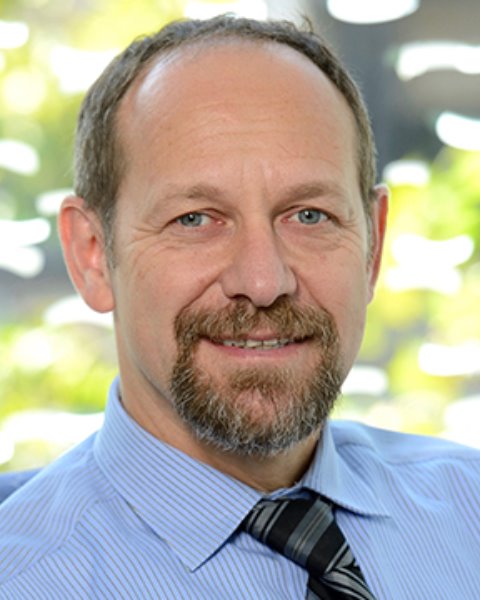
Shirley Ryan AbilityLab
Pablo Celnik, MD, is a physiatrist, physician-scientist and internationally recognized leader in rehabilitation and academic medicine. Dr. Celnik embraces an integrated approach to medicine — one that encompasses the latest in research and technological advances and the best care to make patients better. Dr. Celnik is well-published in highly regarded peer-reviewed journals, has been the recipient of countless prestigious awards and has been inducted into the National Academy of Medicine.
Since Oct. 2023, he has served as Chief Executive Officer of Shirley Ryan AbilityLab, the global leader in physical medicine and rehabilitation for adults and children with the most severe, complex conditions. As CEO, he oversees the organization’s vast clinical and research enterprise, including 2,500 employees, more than 30 sites of care, and affiliates and alliances around the world. Previously, he served as Physiatrist-in-Chief and Chair of the Department of Physical Medicine and Rehabilitation (PM&R) at Johns Hopkins University, leading the rehabilitation service line. His additional responsibilities included serving as Director of the Human Brain Physiology Lab; Director of the Precision Medicine Center of Excellence in Rehabilitation and Co-director of the Sheikh Khalifa Stroke Institute — all at the Johns Hopkins School of Medicine.
A native of Argentina, he received his medical degree from the University of Buenos Aires Faculty of Medical Sciences. He completed his residency training in neurology in Argentina and a fellowship in neurological rehabilitation at the University of Maryland. He also completed two research fellowships at the NIH’s National Institute of Neurological Disorders and Stroke. In 2000, he entered the PM&R residency program at Johns Hopkins, where he ultimately was appointed Chief Resident. For 20 years, he served on the Johns Hopkins faculty in the PM&R, neurology and neuroscience departments.
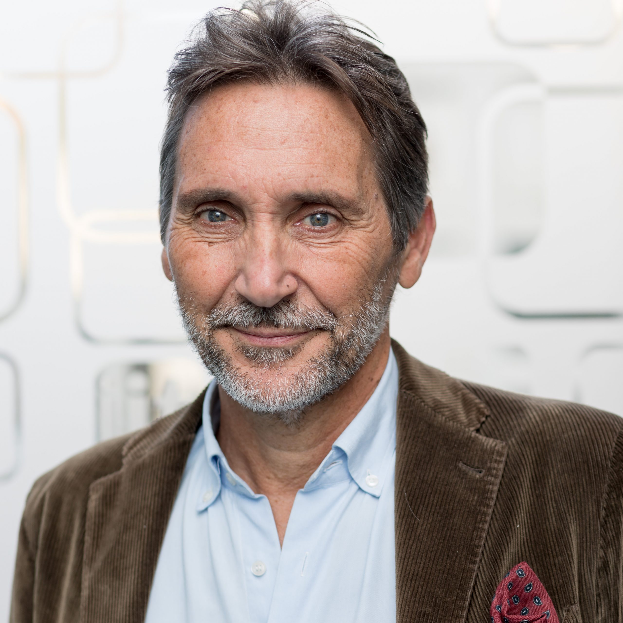
University of Pisa, Italy
In collaboration with Dario Farina and Oskar Aszmann
Antonio Bicchi is a scientist interested in robotics and intelligent machines for human use. He received his Ph.D. from the Alma Mater Studiorum – University of Bologna, and was a postdoctoral fellow at MIT AI Lab, before becoming the first Professor of Robotics at the University of Pisa. In 2009 he founded the Soft Robotics Laboratory at the Italian Institute of Technology in Genoa. Since 2013 he cooperates with Arizona State University as adjunct Professor. He has coordinated many international projects, including four grants from the European Research Council (ERC).
He has authored over 500 scientific papers cited more than 25,000 times. His main contributions are in the field of analysis of grasping and manipulation, the design of soft and variable stiffness hands and limbs, and their control for both robotics and prosthetic applications. He supervised over 70 doctoral students and more than 20 postdocs, most of whom are now professors in universities and international research centers, or have launched their own spin-off companies. His students have received prestigious awards, including four first prizes and two nominations for the best Ph.D. Thesis on Robotics and Haptics subjects. He is a Fellow of IEEE since 2005, and the recipient of the 2018 Saridis Leadership Award.
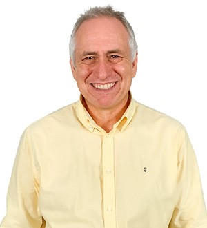
Instituto de Biomecánica de Valencia, Spain
In collaboration with Rosa Porcar-Seder
Abstract: We’ll present the results of a series of experiments conducted to explore new avenues to the analysis of human movements through the scanning of shapes in motion, also known as 4D scanning. 4D scans can provide trajectories of points on the surface of human bodies, with unique identifiers that can be associated with anatomical landmarks without having to instrument the participants. It has been proved that those data can be used to analyze the kinematics of movement, with differences from marker-based systems comparable to inter-examiner errors. In meshes with nearly 50,000 vertices, it is possible to represent anatomical landmarks using dense distributions of points over the target body regions and perform an analysis of gait kinematics with Bayesian statistics. Other ways of measuring the orientation of body segments are possible without having to identify specific landmarks that may be difficult to locate, such as tracking the rotation of contours of body sections with characteristic shapes.
4D scans can be also the source of metrics based on the shape and topology of body segments. Body segment inertial parameters, measured directly from the geometry of the segmented mesh at each instant, can be used to improve joint moment estimation through inverse dynamic analysis in situations where the inertial effect of body masses is significant, in neuromusculoskeletal models this implies reduction of the residuals. Kinematic alterations and asymmetric patterns in soft tissue deformation between the right and left legs have also been found at different instants of the gait cycle, and related to differences in muscle activation, consistent with clinical assessments of people with neurological disorders.
Dr. Juan-Manuel Belda-Lois holds a Ph.D. in Physics from the Universitat Politècnica de València. His Ph. D. thesis was “Biomechanical Principles for Tremor Suppression through Orthotic Means”. He has been working as a researcher at the Instituto de Biomecánica de Valencia since 1995. He is an associate professor in the Master’s in Biomedical Engineering and the Degree’s in Biomedical Engineering programs at the Universitat Politècnica de València. His work is primarily focused on the development and research of assistive products for persons with disabilities, as well as in the field of Rehabilitation Robotics. He has extensive experience in the development and assessment of exoskeletons for rehabilitation and occupational use. Juan Manuel Belda has participated in European projects under the FP5, FP6, FP7, and H2020 programs (including ABC, TREMOR, BETTER, and SUaaVE).
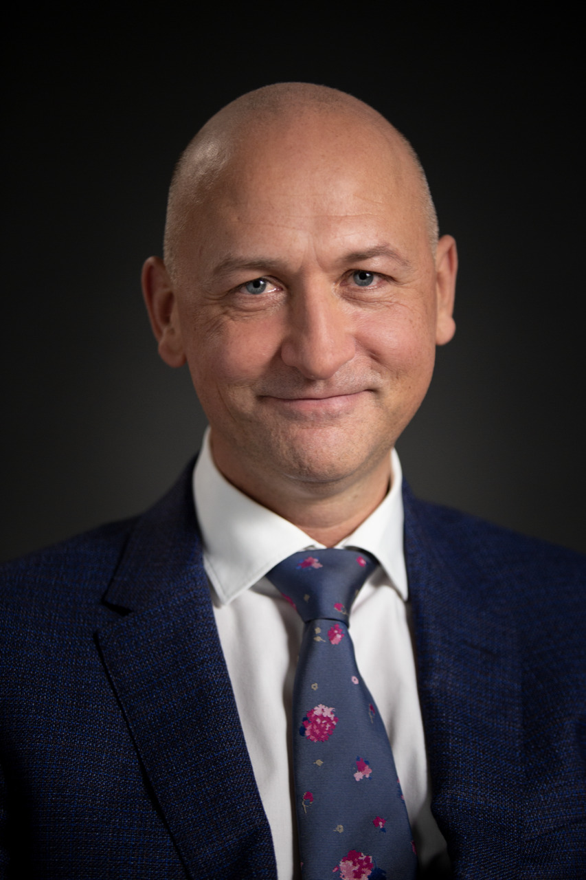
Medical University of Vienna, Austria
In collaboration with Dario Farina and Antonio Bicchi
Prof. Oskar C. Aszmann, born in Vienna, Austria. After a two year excursion into philosophy and biology Dr. Aszmann finished Medical School at the medical faculty of the University of Vienna (1994). He then went on to the Johns Hopkins University in Baltimore, Maryland where he learned the trade of peripheral nerve surgery from Prof. Lee Dellon and the basic science of peripheral nerve regeneration from Prof. Thomas Brushart. In 1998 he joined the Division of Plastic Surgery in Vienna where he was promoted the position of Associate Professor of Plastic and Reconstructive Surgery. Since 2006 he has entered a close collaboration with the company Otto Bock to explore the possibilities and limits of bionic reconstruction which has led to the establishment of a partly private/government funded Center for Extremity Reconstruction and Rehabilitation in 2012. This Center is being headed by Prof. Aszmann and has at its core interest the reconstruction and rehabilitation of patients with impaired extremity function. This goal is accomplished with a wide variety of surgical techniques of neuromuscular reconstruction alone or in combination with complex mechatronic devices. 2020 he has been given the position of Full Professor at the newly founded Department of Plastic and Reconstructive Surgery.
His research focuses on all aspects of reconstructive surgery, both from a clinical but also from a basic research perspective. This has precipitated in different textbook chapters and is being published both in top journals of his field but also larger audience periodicals such as The Lancet and Science, various Nature Group Periodicals and very recently The New England Journal of Medicine. For his accomplishments in this field and his care for patients with complex extremity injuries he was awarded by the Royal Society of Medicine, London twice and received the Hans Anderl Award- the most prestigious research prize awarded by the European Association for Plastic and Reconstructive Surgery for continued excellence in Plastic Surgery Research, the prestigious Houska Award for excellency in public-private partnership and most recently the Christian Doppler Prize for Research and Innovation in September 2020. He serves in the board of directors of several national und international scientific societies and is in the editorial board of several international Journals.
He has received numerous national and international research grants among these, from the Austrian Research Agency (FWF), the Christian Doppler Research Foundation and the European Research Council (ERC) with a sum total of more than 8 Mio€.
